 Fig.1: Smads Downstream Signal Pathway
2015 - present Research at Temple University
Smads are intracellular proteins that transduce extracellular signals from transforming growth factor beta( TGF-beta) ligands to the nucleus where they activate downstream gene transcription as transcription factors (Fig.1). Nuclear Pore Complexes (NPCs) are large protein complexes that cross the nuclear envelope, which allow the transport of molecules across the nuclear envelope.
The different distribution of Smad protein between the absence and presence of transforming growth factor beta has been reported. At presence of TGF-beta, Smad4s strongly concentrate within the nuclear, while no inducing of TGF-beta, Smad4s distribute equally in both of nuclear and cytoplasm. One of hypothesis of mechanism by which Smad4s concentrate within nucleosm is the DNA rentation .
However, the mechanism of Smads transportation through the Nuclear Pore Complexes is still unclear. So research in this mechanism is significant to have a detailed picture of how Smads import and export through NPCs.
Now I work in lab of Dr. Weidong Yang focusing on exploring the mechanism of Nucleocytoplasmic transport of Smads through the Nuclear Pore Complexes. I are confirming whether nuclear retention causes Smad4 inhibition of export by Epi Flurescent Microscopy. We also use SPEED microscope and Single-Molecule Detection In Vivo for exploring differences in the rate, efficiency, time and pathway of Smad4 during export and import through Nuclear Pore Complexes
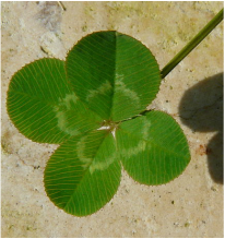 Fig.2 White Clover
2013 - 2014 Research at Northwest A&F University (China)
This research project is research on cell microstructure of V-shape leaf variegation of Trifolium repens.
Trifolium repens, the(Fig.2) white clover (also known as Dutch clover and Ladino), is a herbaceous perennial plant in the bean family Fabaceae native to Europe and central Asia. It has been widely introduced worldwide as a yard crop, and is now also common in most grassy areas of North America and New Zealand.
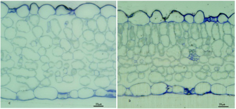 Fig.3 Differences in tissue structure.
To explore the formation mechanism of the white variegation on the white clover leaves, both variegation section and normal section of white clover leaves are gathered as experimental materials. The differences of two sections in pigment content, tissue structure( Fig.3), cell structure and subcellular structure(Fig.4) are studied by following experimental methods including ethanol decolorization, determination of pigment content, semi-thin sectioning technique, electron microscopy.
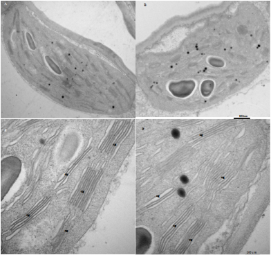 Fig.4 Differences in subcellular structure
Results show that 8 indices of 13 had very significant difference between two sections. The 10 indices include the total chlorophyll content, the thickness of spongy tissue, the thickness of leaf, the ratio of palisade tissue thickness to thickness of leaf, the sparse rate of palisade tissue, the ratio of length to width of palisade cells, the amount of chloroplast grana of palisade cells and the width of chloroplast grana of palisade cells. Moreover,significant differences in structure of tissue between two sections were discovered by comparing microstructures of two sections. Therefore, the differences of two sections mentioned above are very likely to be the cause of formation of variegation on the white clover leaves.
